Automatic MRI image segmentation of the knee
Development of an analysis tool to study knee morphology and biomechanics in vivo
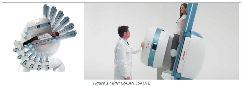
The knee is a complex joint that plays a crucial role in the mobility and functionality of the lower limb. Understanding the morphology and biomechanics of the knee is essential for assessing its health, developing appropriate medical treatments, and contributing to tissue engineering research. As part of an internal study at CARTIGEN, the main objective is to develop an experimental and numerical method for accurately describing the knee's in vivo morphological and biomechanical parameters in healthy subjects. Knee joint morphology can be studied in the majority of MRI scans, but the examinations are not carried out in positions close to everyday life. The GSCAN ESAOTE makes studying the knee in an "ecological" position possible. Such a tool can also be used to study the difference in joint morphology between a horizontal and vertical position.

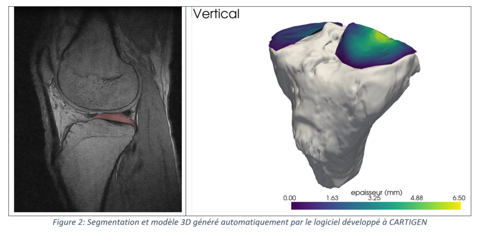
The proposed biomechanical analysis method involves assessing the morphology of articular cartilage in the tibia, femur, and patella in vivo, taking into account the weight-bearing and unweight-bearing positions of the knee. To this end, an automatic knee segmentation tool has been developed within CARTIGEN, with promising results. The similarity indices (DICE) obtained are of the order of 97% for the tibia and 87% for articular cartilage, demonstrating the effectiveness of the method.

In addition to the analysis of cartilage morphology, an in-depth study will be undertaken to assess the relative mobility of the menisci and the anterior and posterior cruciate ligaments during knee flexion movements. The crescent-shaped menisci and cruciate ligaments play an essential role in knee stability and functionality. By assessing their mobility during flexion movements, we can better understand their interaction and contribution to knee biomechanics
The methodology developed in this study will also enable us to assess the impact of subject weight, different types of surgery (meniscectomy, ligament rupture), and medical devices such as knee orthoses on the mechanical loading of the joint. By understanding how surgery or medical devices modify the mechanical loading of the knee, we'll be able to better assess their effectiveness in different clinical contexts.
Finally, the methodology developed in this study also aims to assess cartilage's in vivo mechanical properties. These data are essential for tissue engineering research, as they provide a better understanding of the mechanical characteristics of natural cartilage, and can be passed on to tissue engineering experts for the development of effective cartilage substitutes.
