"Tissue engineering" platform
Devices available
MicroFAB-3D
MicroFAB-3D is an ultra-high-resolution 3D printing system based on direct laser writing technology using two-photon polymerization. With a minimum size of 0.2 microns, microFAB-3D opens up new perspectives in the fields of microfluidics, micro-optics, cell culture, micro-robotics and meta-materials.
BIO X
Cellink's BIO X bioprinter enables 3D bioprinting with up to three print heads, providing multi-material and multi-cell flexibility for small- and large-scale tissue engineering.
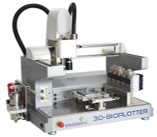
Bioplotter 3D
EnvisionTEC's Bioplotter 3D is one of the most experienced bioprinters on the market, backed by unprecedented research and development. These process open-source biomaterials, using air or mechanical pressure to a syringe for computer-aided tissue engineering, scaffolds to create tissues, organs, physical structures and more. This bioprinter is designed for use in a sterile biosafety cabinet and meets clinical trial standards.
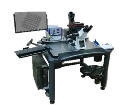
LEICA TCS SP8 DLS confocal microscope
Fully motorized, LAS-controlled inverted confocal microscope for epifluorescence and transmission. UV-optimized light path. Fluorescence (LASERs 355, 405, Argon, DPSS 561 and HeNe 633) and brightfield/DIC. Multi-dimensional acquisition.
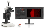
THUNDER Imager Model Organism
THUNDER Imager Model Organism is a stereomicroscope for imaging model organisms used in cell and developmental biology, such as Drosophila, nematodes, zebrafish, mouse models, etc... It provides a clear view of even the deepest details of an intact sample, in real time, eliminating the blurring inherent in wide-field imaging.
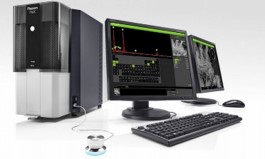
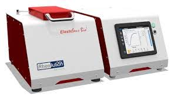
Phenom Pro Desktop SEM
The Phenom Pro Desktop SEM is a desktop scanning electron microscope (SEM) and can be used to ease the burden of routine analysis of common samples from basic SEM instruments. The instrument's configuration and sample loading mechanism guarantee rapid imaging and minimal set-up time between experiments. Its high stability and small size enable the instrument to be used in virtually any laboratory environment.
ElastoSens Bio
Rheolution's ElastoSens Bio enables non-destructive mechanical testing of flexible biomaterials. This tabletop instrument enables the evolution of viscoelasticity to be characterized precisely and in real time, without contact and without destroying the sample.
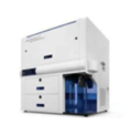
Symphony A3
The Symphony A3 cytometer is a powerful analytical tool that enables users to identify and analyze distinctive phenotypes in heterogeneous populations. Featuring 5 lasers with 28 color parameters, it is dedicated to complex multi-parametric studies.
The most sensitive and rapid system on the market, it enables users to search for rare events. The configuration of the equipment allows the use of different wavelengths and traditional and/or unique powers.
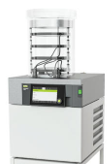
Lyophylisateur L-200
L-200 compact freeze-dryer, the first freeze-dryer with Infinite-Control™, offers unrivalled automation in sublimation. Easily create and run methods, log data, record real-time graphs and interact wherever you are using your mobile devices.
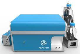
NanoAnalyzer NanoFCM
The NanoAnalyzer NanoFCM is a flow cytometer specifically designed to analyze nano-sized particles such as extracellular vesicles (EVs), viruses and nanoparticles, based on light scattering and fluorescence for the detection of single virions or EVs down to 40 nm.
The development of a pipeline approach based on flow nano-cytometry will enable multi-parametric quantitative analysis of nanoparticles, which is very important for the study of their biological functions.
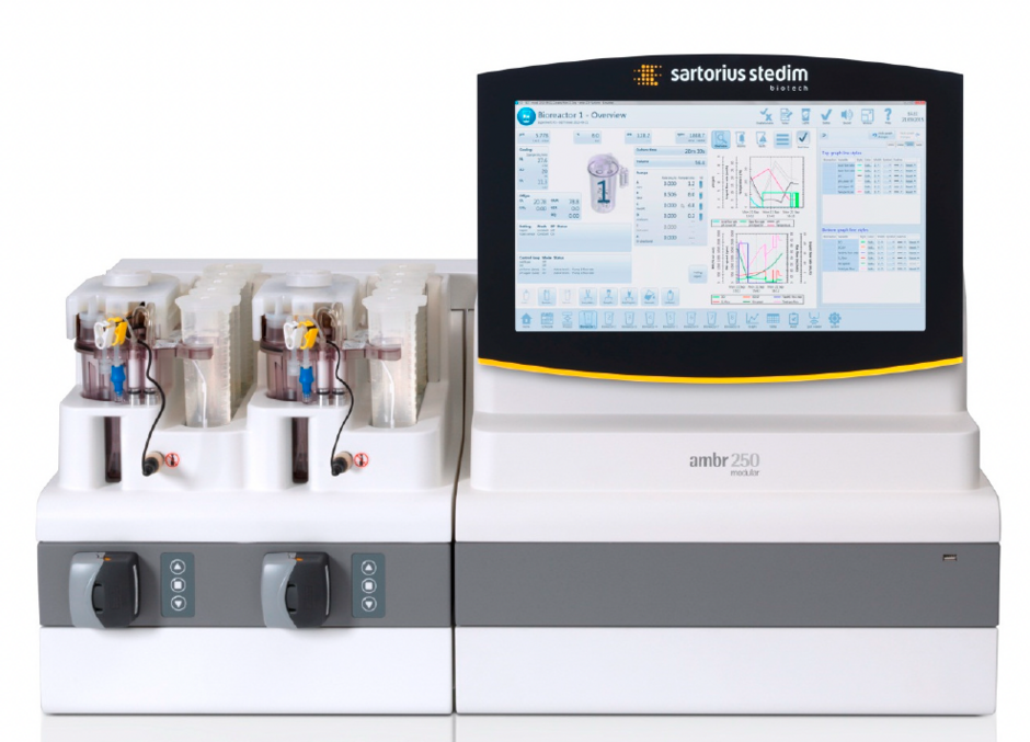
Ambr® 250 modular
Ambr® 250 modular from Sartorius is an innovative new high-performance benchtop bioreactor system for parallel microbial or cell culture in single-use vessels from 100 to 250 ml. The system uses advanced stirred-tank bioreactor technology. The system comprises a series of benchtop modules for running 1 to 8 bioreactors in parallel, plus a control module with intuitive system software accessible via a user interface screen.
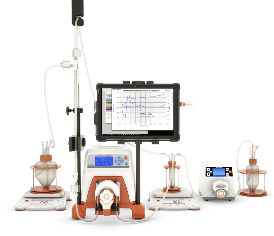
Tangential flow filtration
The KR2i tangential flow filtration (TFF) system [KrosFlo® Research 2 i] is the ideal automated pumping system for processes from 1ml to 10L.
The KR2i is the ideal system for microfiltration and ultrafiltration in small volumes and on the research and development scale, thanks to its small footprint and versatility. TFF systems offer numerous advantages over traditional cross-flow membrane systems. The hollow-fiber (HF) membrane modules and cross-flow filtration system enable faster, gentler separation, helping to avoid membrane fouling and maximize product recovery.
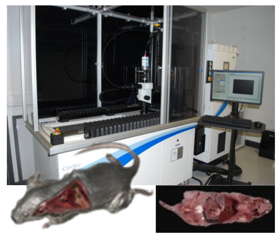
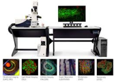
CryoViz
The CryoViz imaging system has been developed for 3D microscopic imaging of mice or whole organs. It is possible to detect and spatially map almost every stem cell in an organ or mouse, offering a unique homing/engraftment assessment capability. CryoViz imaging is ideal for answering the almost ubiquitous question in stem cell therapies and cancer applications: "Where did these cells go?". Moreover, thanks to differentiation reporter genes, it is possible to map cells that have undergone differentiation in vivo.
By alternating cutting and imaging, the system acquires volumes of 3D color and fluorescence images from sequential images of the face of the tissue block. Multiscale volume rendering provides views of anatomy as well as molecular fluorescence showing the location of molecules and/or cells. This technology bridges the gap between whole-animal in vivo imaging and histology, enabling a mouse to be imaged along the entire mouse -> organ -> tissue structure -> cell -> subcellular domain continuum.